Dichroic filters in microscopy are vital for science. They help study cells, tissues, and tiny life forms. Scientists use them to see fluorescent molecules clearly, making important findings. These filters let some light through while blocking others, making images sharper. In biology and medicine, they reveal secrets about diseases and cures. This intro dives into why dichroic filter microscopy matters.
Definition of Dichroic Filter Fluorescence Microscopy
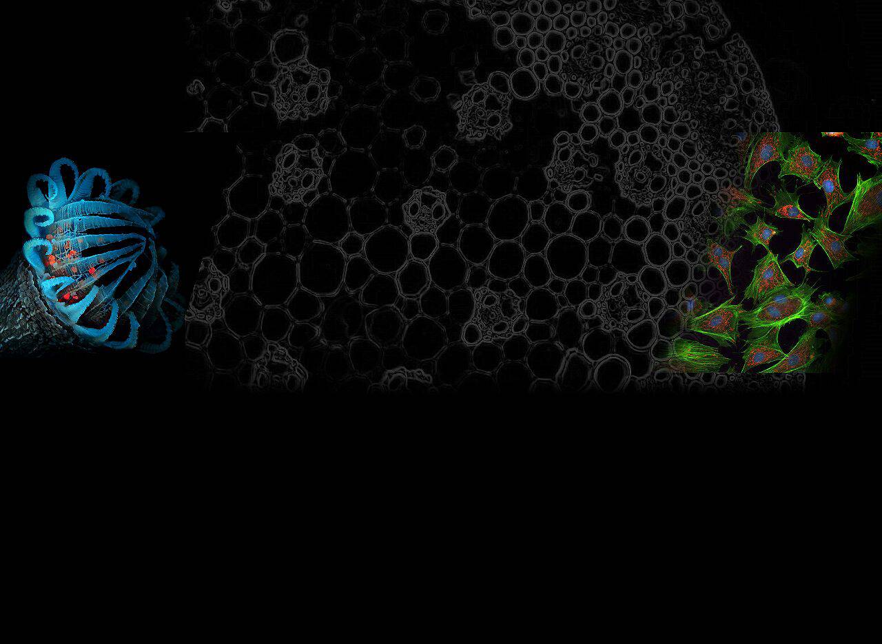
Dichroic Filter Fluorescence Microscopy helps us see tiny things in biology and materials. It uses special filters, like traffic cops for light. These filters let some light through and reflect other colors. When light hits a sample, it makes some molecules glow in different colors. The filter lets this special light through to the detector. This separation makes images sharp and clear.
What is the function of the dichroic filter in fluorescence microscopy?
The dichroic filter in fluorescence microscopy serves a pivotal role in capturing detailed and vibrant images of microscopic specimens. This specialized filter is like the maestro of the microscope, orchestrating the symphony of light to illuminate and visualize the hidden world of cells, tissues, and molecules. Let’s delve into the function of the dichroic filter and why it’s so vital:
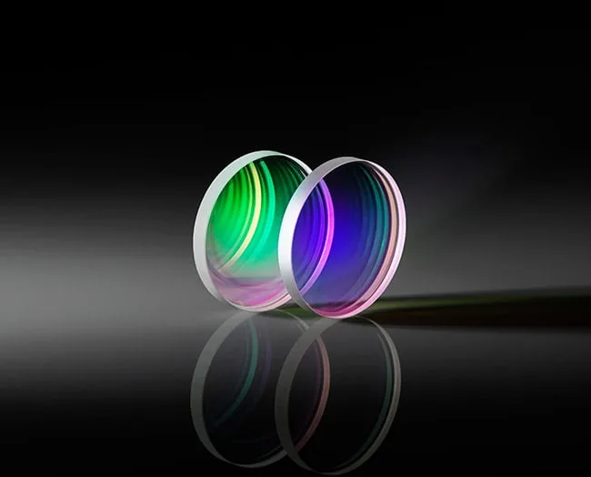
Wavelength Sorting
The primary job of the dichroic filter is to act as a traffic cop for light. It separates incoming light into different wavelengths or colors. This is essential because, in fluorescence microscopy, we want to selectively focus on specific colors of light to get a clear view of our specimen.
Excitation and Emission Light Control
In fluorescence microscopy, there are two types of light: excitation light and emission light. The dichroic filter is like a gatekeeper that decides who gets in and who doesn’t. It reflects the high-energy excitation light towards the specimen while allowing the lower-energy emission light from fluorophores within the specimen to pass through.
Enhanced Contrast
By separating the excitation and emission light, the dichroic filter significantly boosts image contrast. This contrast enhancement makes it easier to spot fluorescent molecules against a dark background, resulting in crisper, more detailed images.
Multicolor Imaging
In many cases, scientists want to visualize multiple fluorescent markers simultaneously. The dichroic filter makes this possible by reflecting one color of excitation light while allowing another to pass through. This enables researchers to study multiple aspects of a specimen at once.
Reduced Photobleaching
Photobleaching is the process where the fluorophores lose their ability to emit light over time. The dichroic filter plays a role in minimizing this issue by ensuring that only the necessary excitation light interacts with the specimen. This means less unnecessary light exposure, which can help extend the lifespan of fluorescent molecules and allow for longer imaging sessions.
Fluorophore Selection
Different fluorophores emit light at different wavelengths. The dichroic filter can be carefully selected to match the emission wavelength of the fluorophore used in the experiment. This ensures that only the specific color of light emitted by the fluorophore is captured, eliminating any interference from other light sources.
High-Resolution Imaging
The dichroic filter contributes to the high resolution of fluorescence microscopy. By controlling which wavelengths of light are used for excitation and detection, it helps achieve sharp, detailed images that are crucial for scientific research and medical diagnostics.
Real-Time Imaging
In live cell or real-time imaging, the dichroic filter’s ability to quickly switch between excitation and emission light paths is crucial. This dynamic function allows researchers to monitor fast-paced biological processes and capture rapid changes in fluorescence.
Customization
Researchers have the flexibility to choose dichroic filters tailored to their specific experimental needs. These filters can be designed to match the fluorophores being used and the microscope’s configuration, ensuring optimal imaging conditions.
Scientific Discovery
Ultimately, the dichroic filter in fluorescence microscopy enables scientists to explore the intricate details of the microscopic world.
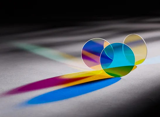
Choosing the Right Dichroic Filter
Selecting the right dichroic filter is like picking the perfect tool for a specific job in fluorescence microscopy. It’s crucial because using the wrong filter can muddy your images and make your research less accurate. Here’s a friendly guide on how to choose the right one:
Know Your Fluorophores
Start by understanding the fluorescent dyes or markers you’re using in your experiment. Each fluorophore emits light at a specific wavelength, so you need a dichroic filter that matches that wavelength.
Microscope Configuration
Consider the setup of your microscope. Different microscopes may require specific filter sizes or types to fit correctly. Check your microscope’s manual or consult with the manufacturer.
Desired Emission Wavelength
Determine the wavelength range of the emitted light you want to capture. Select a dichroic filter that allows this range to pass through while reflecting unwanted wavelengths.
Multicolor Imaging
If you’re doing multicolor imaging, you’ll need to choose filters that work together harmoniously. Ensure that the filters you select won’t cause interference or bleed-over between different fluorophores.
Filter Quality
Invest in high-quality filters. Cheap filters can distort colors and compromise image quality. Look for reputable brands or consult with experts in microscopy.
Application-Specific Filters
Some experiments may require specialized filters, like polarization filters or filters optimized for specific imaging techniques (e.g., FRET or FRAP). Make sure your chosen filter suits your application.
Consult Experts
Don’t hesitate to seek advice from experienced microscopists or colleagues who have expertise in fluorescence microscopy. They can provide valuable insights and recommend suitable filters.
Test and Validate
Before diving into your experiments, test the filter’s performance with your specific fluorophores and conditions. This ensures that your chosen filter will yield accurate and reliable results.