Fluorescence microscopy has become an essential technique for cellular and molecular biology research, providing the ability to visualize structures and dynamics within live and fixed cells. The use of specific optical filters designed for different fluorophores is crucial for maximizing image clarity and reducing background interference. Among the many available fluorophores, DAPI (4′,6-diamidino-2-phenylindole) is one of the most widely used due to its ability to stain DNA and reveal nuclei in blue fluorescence. To achieve the best possible imaging results with DAPI, selecting the correct optical filters is essential. These filters are specifically designed to match the excitation and emission wavelengths of DAPI, ensuring optimal fluorescence detection.
In this article, we will explore the role of DAPI filters, the excitation and emission wavelengths associated with DAPI, its spectrum, and the importance of using the correct DAPI emission wavelength for enhanced imaging.
Understanding DAPI’s Excitation and Emission Characteristics
DAPI is a fluorescent dye that binds to the minor groove of double-stranded DNA, making it a popular choice for nuclear staining in both fixed and live cells. However, for effective fluorescence detection, it’s critical to use appropriate optical filters that match the dye’s excitation and emission spectra.
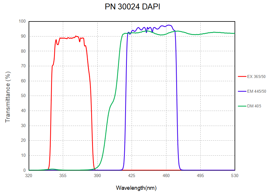
DAPI Excitation Wavelength: The excitation wavelength of DAPI typically falls in the 350-360 nm range. To achieve optimal fluorescence, a UV or violet light source, often from a mercury vapor lamp or a laser, is used to excite the dye. The excitation filter must allow light at these wavelengths to pass through while blocking other unwanted wavelengths, ensuring that only DAPI is excited.
DAPI Emission Wavelength: Upon excitation, DAPI emits light in the 460-470 nm range. This emission wavelength falls in the blue region of the visible spectrum. The emission filter is used to capture this emitted light while blocking out any other light from the excitation source or other fluorophores in multi-color experiments.
The Role of DAPI Filters in Fluorescence Microscopy
In fluorescence microscopy, optical filters are used to isolate specific wavelengths of light, enabling accurate detection of the fluorescence signal from the labeled specimen. When working with DAPI, it is essential to select the appropriate filters for both excitation and emission to ensure that the fluorescence is captured with maximum efficiency and minimal background interference.
DAPI Excitation Filter: The excitation filter is designed to allow light at the specific wavelength that excites DAPI (around 350-360 nm) to pass through, while blocking other wavelengths. It typically has a narrow bandwidth to prevent the overlap of excitation light from other sources, which could lead to background fluorescence and reduced contrast.
DAPI Emission Filter: The emission filter works in conjunction with the excitation filter to capture the emitted blue fluorescence from DAPI after it is excited. The emission filter must allow light in the 460-470 nm range to pass through while blocking any stray excitation light that might still be present. The high specificity of this filter is essential to ensure the clarity and brightness of the fluorescence signal.
DAPI Spectrum and Filter Design
A key feature of DAPI is its distinct fluorescence spectrum, which is characterized by a sharp peak of emission around 461 nm. The DAPI spectrum helps in the design of optical filters that can precisely isolate the desired excitation and emission wavelengths.
DAPI Spectrum: The DAPI spectrum can be viewed as a broad peak that spans approximately from 350 nm (excitation) to 470 nm (emission). The sharp emission peak at 461 nm is crucial for designing the emission filter, which should have a narrow bandpass that specifically isolates light in this range for high-contrast imaging. In contrast, the excitation filter should be chosen to selectively transmit light in the UV range (350-360 nm) without including other wavelengths, such as those emitted by common fluorescence lamps or lasers for other fluorophores.
Filter Design Considerations: The filter sets for DAPI typically consist of a combination of an excitation filter with a peak transmission at around 350-360 nm, a dichroic mirror that reflects the excitation light while allowing emitted light to pass through, and an emission filter that transmits the light around 461 nm. The narrow passbands of the filters minimize the chances of spectral overlap and ensure that only DAPI fluorescence is detected.

Choosing the Right DAPI Optical Filter Set
Selecting the optimal optical filter set for DAPI fluorescence microscopy depends on several factors, including the type of microscope and light source being used, as well as the desired imaging quality. In general, the following considerations should guide the selection of DAPI filters:
Filter Bandwidth: Both the excitation and emission filters should have narrow bandwidths to minimize overlap with the emission spectra of other fluorophores or autofluorescence from the specimen.
Filter Quality: High-quality filters with minimal transmission loss and low autofluorescence are essential for obtaining clear and bright images. This is especially important for experiments requiring high sensitivity and high signal-to-noise ratios.
Multi-Color Imaging: If using DAPI in a multi-color fluorescence experiment, careful attention must be paid to the spectral separation of the filters. Using a filter set with a well-defined passband for DAPI ensures that its fluorescence can be distinguished from other fluorophores, such as GFP or RFP, which may be used in the same experiment.
Applications of DAPI Filters in Fluorescence Microscopy
DAPI filters are particularly useful in several research applications, including:
Examine nuclear morphology, cell division, and apoptosis. Proper optical filters are crucial for achieving clear, high-contrast images of the nuclear structures.
Cell Cycle Analysis: DAPI staining is widely used in flow cytometry and other cell cycle studies to quantify DNA content. Accurate detection of DAPI fluorescence using the right optical filters ensures precise measurements of DNA quantity in cells.
Subcellular Localization: DAPI filters are also used in combination with other fluorescent probes to study the localization of specific proteins within the nucleus. This can be particularly valuable in studies of gene expression, protein trafficking, and nuclear processes.
Conclusion
In fluorescence microscopy, the proper use of optical filters is essential for maximizing the sensitivity and clarity of fluorescence signals, particularly when working with dyes like DAPI. Understanding the excitation and emission wavelengths of DAPI, as well as its spectrum, is key to designing effective filter sets that isolate the correct wavelengths for imaging. With the right DAPI emission wavelength filters, researchers can achieve high-resolution, high-contrast images of DNA and cellular structures, facilitating groundbreaking discoveries in cellular and molecular biology.
