Fluorescence microscopy has revolutionized the way we observe and understand the microscopic world, enabling researchers to explore the intricacies of cellular structures and molecular interactions.
Among the various fluorophores available, Tritc (Tetramethylrhodamine Isothiocyanate) stands out for its unique features and exceptional performance in fluorescence imaging.
Today we will explore the features and performance of Tritc wavelength in fluorescence microscopy. Let’s go to read.

Key Feature of Tritc Wavelength in Fluorescence Microscopy
TRITC (Tetramethylrhodamine Isothiocyanate) wavelength in fluorescence microscopy offers several distinct features that contribute to its widespread use in the field. Here are some key features of Tritc wavelength:
Distinctive Red-Orange Fluorescence
Tritc emits a distinct red-orange fluorescence when excited, making it easily distinguishable from other fluorophores. This characteristic color allows researchers to specifically label and visualize target structures within biological samples, providing a clear and identifiable signal.
Enhanced Sensitivity
Tritc is known for its high sensitivity, making it particularly effective in capturing weak fluorescence signals. This feature is crucial for imaging applications where the target molecules may be present in low concentrations or exhibit faint fluorescence. The enhanced sensitivity of Tritc contributes to improved image quality and the detection of subtle biological processes.
Compatibility for Multicolor Imaging
One of the standout features of Tritc is its compatibility with other fluorophores, enabling researchers to perform multicolor imaging experiments. By combining Tritc with fluorophores emitting at different wavelengths.
It becomes possible to simultaneously visualize multiple cellular components or molecular events within the same sample. This versatility is essential for gaining a comprehensive understanding of complex biological systems.
Photostability
Tritc exhibits excellent photostability, meaning that it resists fading or photobleaching during prolonged exposure to excitation light. This feature is crucial for time-lapse imaging and extended experiments, ensuring that, the fluorescence signal remains robust throughout the study. The photostability of Tritc contributes to the reliability of fluorescence microscopy results.
Reduced Photobleaching
Compared to some other fluorophores, Tritc demonstrates reduced photobleaching. Photobleaching occurs when fluorophores lose their fluorescence intensity over time due to exposure to light.

Performance of Tritc wavelength in Fluorescence Microscopy
Tetramethylrhodamine isothiocyanate (TRITC) is a commonly used fluorochrome in fluorescence microscopy. TRITC is excited by light in the blue-to-green range (around 488 nm), and it emits fluorescence in the orange-to-red range (around 580 nm). Here are some key points regarding the performance of TRITC in fluorescence microscopy.
Excitation and Emission Wavelengths
Excitation Wavelength: TRITC is excited most efficiently in the range of 510-560 nm, with a peak excitation at around 550 nm. As well as the emitted light is typically in the range of 570-650 nm, with a peak emission of around 580 nm.
Fluorescence Brightness
TRITC is known for its relatively bright fluorescence signal, making it suitable for imaging applications where a strong signal is crucial.
Photostability
TRITC generally exhibits good photostability, meaning it can withstand prolonged exposure to excitation light without significant loss of fluorescence intensity. This is important for time-lapse imaging and other applications where extended exposure times are necessary.
Spectral Overlap
One consideration when using TRITC is its spectral overlap with other fluorochromes. If you are using multiple fluorochromes in a single experiment, it’s important to choose fluorochromes with distinct excitation and emission spectra to avoid spectral bleed-through and ensure accurate multicolor imaging.
Filter Sets
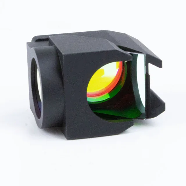
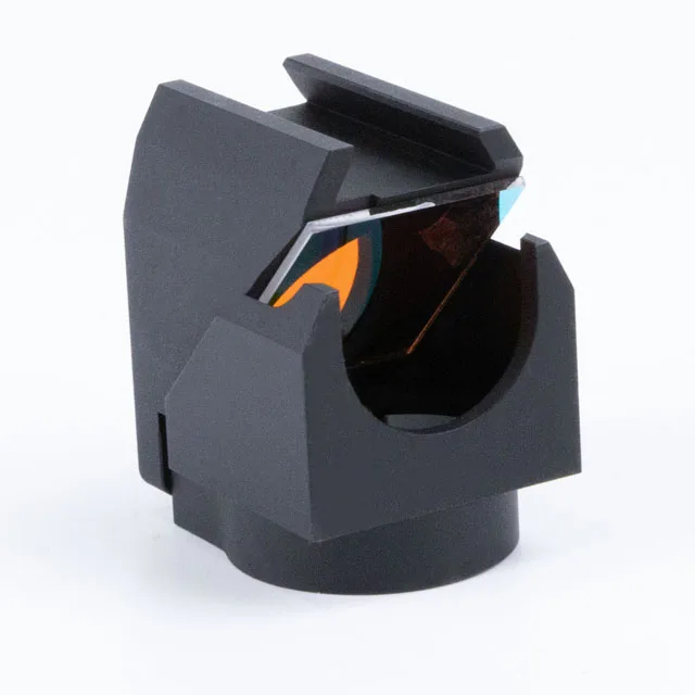
To effectively use TRITC in fluorescence microscopy, appropriate filter sets are required. This typically includes an excitation filter that allows light in the range of 510-560 nm to pass through and an emission filter that transmits light in the 570-650 nm range.
Applications
TRITC is commonly used in immunofluorescence staining, where it can be conjugated to antibodies to label specific cellular components.
Considerations
When working with TRITC, consider factors such as the intensity of the fluorescence signal, potential photobleaching, and compatibility with other fluorochromes used in the experiment.
Keep in mind that the performance of TRITC can also depend on the specific microscope setup, the quality of filters and light sources, and other experimental conditions. Always refer to the manufacturer’s recommendations and optimize your microscopy settings for the best results with TRITC.
Final thought
Tritc wavelength in fluorescence microscopy emerges as a powerhouse, offering enhanced sensitivity, versatility in multicolor imaging, excellent photostability, and minimized photobleaching.
