Introduction
Technological developments have been essential in solving the riddles of life in the dynamic field of biological research. Fluorescence microscopy using a DAPI filter is one such revolutionary invention. This cutting-edge method has completely changed how scientists view and analyse biological samples, opening up new research directions and deepening our understanding of living things. We shall examine the many ways that DAPI filter fluorescence microscopy has influenced biological research in this extensive article, from its principles to its wide range of applications.
How Does DAPI Filter Fluorescence Microscopy Revolutionize Biological Research?
We will go into the fundamentals of DAPI filter fluorescence microscopy in this section to better comprehend how it has revolutionised biological research.
Understanding Fluorescence Microscopy
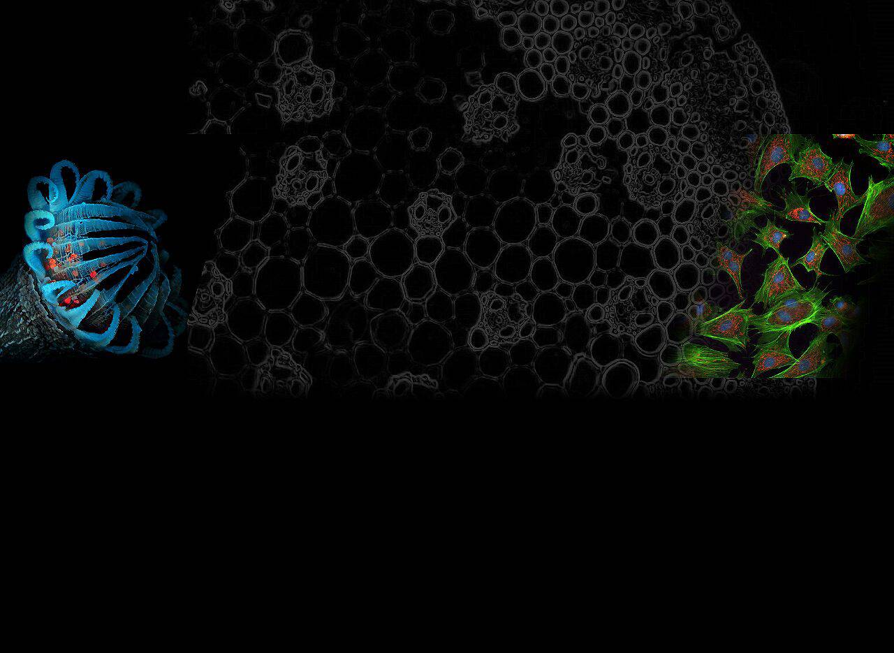
An imaging method called fluorescence microscopy uses fluorescence to examine biological material. Fluorophores, chemicals that can emit light when activated by particular light wavelengths, are used in this process. Scientists may visualise and investigate cellular processes in real-time by labelling biological structures with fluorescent markers, yielding important insights into a variety of biological phenomena.
The Significance of DAPI in Fluorescence Microscopy
Popular fluorophore DAPI (4′,6-diamidino-2-phenylindole) is well known for its remarkable DNA-binding abilities. The characteristic blue fluorescence that DAPI generates after binding to DNA allows scientists to see the cell nucleus with unmatched clarity. Due to its distinctive properties, DAPI has become a crucial tool in fluorescence microscopy, particularly for research on cell division, nuclear morphology, and chromatin organisation.
Advantages of DAPI Filter Fluorescence Microscopy
High Resolution Imaging: Scientists can obtain high-resolution images using DAPI filter fluorescence microscopy, which captures delicate features of cellular structures with outstanding clarity. This has significantly increased our understanding of cellular biology.
Specific DNA Labeling: The remarkable specificity of DAPI’s DNA binding properties allows for the targeted labelling of cell nuclei. The precision of observations is improved and background noise is reduced by this specificity.
Live Cell Imaging: Live cell imaging using DAPI filter fluorescence microscopy enables researchers to track dynamic cellular processes in real-time, leading to ground-breaking discoveries.
Multiplexing Capabilities: DAPI can be used in concert with other fluorophores to mark numerous cellular structures at once and explore intricate interactions within the cell.
Quantitative Analysis: Cell cycle studies and genetic research are made possible by the quantitative analysis of DNA content made possible by DAPI filter fluorescence microscopy.
Applications in Biological Research
Numerous biological research fields have found widespread use for DAPI filter fluorescence microscopy. The following are some of the principal areas where it has had a big impact:
Cell Biology
Cell Cycle Studies: DAPI is frequently used to examine the cell cycle and offers insightful data on cell replication and division.
Apoptosis Research: DAPI helps identify nuclear condensation, a sign of apoptosis, and aids in the investigation of processes involved in programmed cell death.
Cell Nucleus Analysis: The exact nuclear morphology study made possible by DAPI aids in the detection of aberrant cellular states.
Developmental Biology
Embryogenesis Studies: When examining embryonic development, DAPI filter fluorescence microscopy is crucial because it sheds light on how tissues grow and differentiate.
Germ Cell Research: Studies pertaining to fertility can benefit from the visualisation of germ cell growth and localization made possible by DAPI.
Genetics and Genomics
Chromatin Organization: Understanding chromatin shape and organisation is essential for comprehending gene regulation and epigenetic changes, and DAPI facilitates this research.
Fluorescence In Situ Hybridization (FISH): In order to see specific DNA sequences and aid in genomic research, DAPI is frequently used in conjunction with FISH methods.
Neurobiology
Neuronal Imaging: The complicated structure and connectivity of neurons are investigated using DAPI filter fluorescence microscopy, increasing our understanding of the nervous system.
Neurodegenerative Diseases: DAPI helps investigate how neurodegenerative illnesses affect neuronal nuclei, providing important information into how the diseases operate.
Impact on Scientific Discoveries
The impact of DAPI filter fluorescence microscopy on scientific breakthroughs is immeasurable. Its contribution to the advancement of numerous biological research domains cannot be underestimated. Some significant effects include:
Improved Disease Understanding: Our understanding of numerous diseases has expanded thanks to DAPI filter fluorescence microscopy, opening the door for interventions and medicines that are specifically targeted.
Drug Development: Drug development processes have been sped up because to insights gained using DAPI filter fluorescence microscopy that gave researchers a thorough understanding of how cells interact and how drugs affect them.
Biotechnology Advancements: The development of DAPI has aided in the development of genetic engineering and gene editing methods in biotechnology.
Advances in Neuroscience: Neurological advancements have resulted from the understanding of the intricacies of the brain through the use of DAPI filter fluorescence microscopy.
Contributions to Developmental Biology: The study of embryonic development using DAPI has benefited tissue engineering and regenerative medicine.

FAQs
FAQ 1: What is DAPI filter fluorescence microscopy?
An imaging method called DAPI filter fluorescence microscopy makes use of the fluorophore DAPI to make DNA in biological samples visible. When DAPI binds to DNA, it releases blue fluorescence, making it possible to image cell nuclei precisely.
FAQ 2: What is the procedure for DAPI filter fluorescence microscopy?
UV light is used to excite the DAPI fluorophore in DAPI filter fluorescence microscopy. The blue fluorescence that is released when DAPI attaches to DNA in the cell nucleus is photographed and seen under a microscope.
FAQ 3: What advantages can DAPI filter fluorescence microscopy offer?
High-resolution imaging, precise DNA labelling, live cell imaging capabilities, multiplexing options, and quantitative analysis of DNA content are just a few advantages of using DAPI filter fluorescence microscopy.
FAQ 4: Which biological research fields frequently employ DAPI filter fluorescence microscopy?
Research in cell biology, developmental biology, genetics, genomics, and neurology uses DAPI filter fluorescence microscopy extensively.
FAQ 5: How has scientific research been affected by DAPI filter fluorescence microscopy?
DAPI filter fluorescence microscopy has significantly advanced our understanding of diseases, contributed to drug development, facilitated biotechnological advancements, and made groundbreaking contributions to neuroscience and developmental biology.
FAQ 6: Can live cell imaging be done with DAPI filter fluorescence microscopy?
Yes, live cell imaging using DAPI filter fluorescence microscopy is possible, allowing researchers to monitor and analyse dynamic cellular activities in real-time.
Conclusion
DAPI filter fluorescence microscopy has become a potent and crucial technique in contemporary biological research, to sum up. Numerous scientific developments and discoveries have been made possible by its capacity to visualise DNA with high sensitivity and resolution across a variety of disciplines. DAPI filter fluorescence microscopy continues to spur innovation and encourage a greater understanding of life itself, from illuminating the mysteries of cell biology to dissecting the complexity of the brain.