Fluorophores are essential components in the field of molecular imaging, playing a crucial role in visualizing and tracking biological molecules within cells and tissues.
They work in tandem with fluorescence filters to ensure that only specific wavelengths of light are absorbed and emitted, enhancing the clarity and specificity of the imaging process.
What is a Fluorophore?
1. Basic Definition and How They Work
A fluorophore is a fluorescent chemical compound that can re-emit light upon light excitation. When exposed to specific wavelengths of light, fluorophores absorb photons and, with the aid of fluorescence filters, re-emit them at longer wavelengths, producing a visible fluorescence.
This unique property, enhanced by the selective capability of fluorescence filters, makes fluorophores invaluable tools for labeling and imaging biological structures, allowing for precise visualization and tracking of biological processes within cells and tissues.
2. Types of Fluorophores
There are various types of fluorophores, including organic dyes, fluorescent proteins, and semiconductor nanocrystals known as quantum dots. Each type has distinct characteristics that make it suitable for different applications in molecular imaging.
Why Fluorophores are Important in Science
1. The Role of Fluorescence in Research
Fluorophores play a pivotal role in scientific research by enabling the visualization of cellular structures, protein interactions, and dynamic processes within living organisms. Their ability to emit light allows researchers to track the movement and behavior of specific molecules in real-time.
2. Examples of Fluorophore Applications
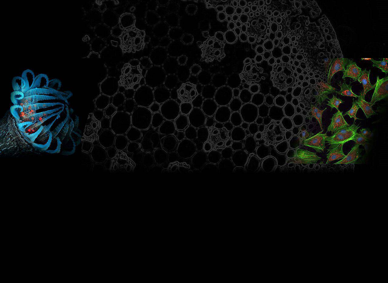
- Cell Labeling: Fluorophores are used to label specific organelles or proteins within cells, allowing scientists to observe their localization and dynamics.
- Molecular Probes: They serve as molecular probes for studying DNA, RNA, and other biomolecules, aiding in the understanding of genetic processes.
The Role of Fluorophores in Molecular Imaging
Fluorophores play a pivotal role in enhancing various imaging techniques, allowing scientists to visualize and study biological structures at the molecular level.
1. How Fluorophores Enhance Imaging Techniques
Fluorescence Microscopy
Fluorescence microscopy utilizes fluorophores to visualize specific molecules within biological samples. By labeling target structures with fluorescent markers, researchers can observe the intricate details of cells and tissues with exceptional clarity.
Confocal Microscopy
In confocal microscopy, fluorophores are used to achieve high-resolution imaging by eliminating out-of-focus light. This technique enables the reconstruction of three-dimensional images, providing valuable insights into the spatial organization of cellular components.
2. Fluorophores in Action: Real-World Examples
Tracking Cellular Processes
Fluorophores are employed to track dynamic cellular processes such as cell division, migration, and signaling events. By labeling specific molecules with fluorescent markers, researchers can monitor these processes in real-time, gaining a deeper understanding of cellular behavior.
Diagnosing Diseases
In medical diagnostics, fluorophores are utilized for imaging techniques that aid in disease diagnosis. For instance, they can be used to detect specific biomarkers associated with diseases such as cancer, enabling early detection and precise localization of pathological conditions within the body.
Choosing the Right Fluorophore for Your Needs
When selecting a fluorophore for molecular imaging, several factors should be carefully considered to ensure optimal results and compatibility with the intended application.
1. Factors to Consider When Selecting a Fluorophore
Wavelength and Brightness
The choice of fluorophore is heavily influenced by its excitation and emission wavelengths, which determine the specific colors it can emit. Additionally, brightness plays a crucial role in signal detection, as brighter fluorophores enhance the visibility of labeled structures, especially in low-light conditions.
Stability and Compatibility
The stability of the fluorophore ensures that the fluorescence signal remains consistent throughout the observation period. In addition, compatibility with sample preparation methods and imaging equipment is essential in order to avoid possible interference or artifacts during the imaging process.
2. Popular Fluorophores and Their Uses
Organic Dyes vs Quantum Dots
Organic dyes are versatile fluorophores known for their broad range of excitation and emission wavelengths, making them suitable for multicolor imaging applications. On the other hand, quantum dots, semiconductor nanocrystals with tunable emission properties, offer exceptional brightness and photostability, making them ideal for long-term imaging studies.
Biological Fluorophores
Biological organisms naturally produce certain proteins or compounds that exhibit fluorescent properties. These biological fluorophores, such as green fluorescent protein (GFP), have revolutionized molecular imaging by enabling non-invasive labeling of cellular structures within living organisms.
Practical Steps in Using Fluorophores for Molecular Imaging

Once the appropriate fluorophore has been selected for molecular imaging, the next crucial steps involve preparing the sample with the chosen fluorophore and capturing as well as analyzing fluorescent images.
1. Preparing Your Sample with Fluorophores
Labeling Techniques
The process of labeling samples with fluorophores requires careful consideration of the specific biological structures or molecules to be visualized. Various labeling techniques, such as direct conjugation and indirect immunofluorescence, can be employed based on the nature of the sample and the desired imaging outcome. Each technique offers distinct advantages and considerations, influencing the choice of method for successful fluorophore labeling.
Avoiding Common Pitfalls
When preparing samples with fluorophores, it is essential to be mindful of potential pitfalls that could compromise imaging results. Factors such as non-specific binding, photobleaching, and inadequate washing steps can impact the quality and specificity of fluorescent labeling.
By employing proper controls, optimizing labeling conditions, and validating imaging protocols, researchers can mitigate these common pitfalls and ensure reliable fluorescence signals.
2. Capturing and Analyzing Fluorescent Images
Setting Up the Imaging Equipment
Capturing high-quality fluorescence images requires meticulous attention to the setup of the imaging equipment. Parameters such as excitation intensity, emission filters, and detector settings must be optimized to maximize the signal-to-noise ratio while minimizing background fluorescence. It is also important to maintain consistent imaging conditions (e.g., temperature and humidity levels) when using fluorophores in molecular imaging.
Interpreting the Results
Upon capturing fluorescent images, thorough analysis and interpretation are vital for extracting meaningful information from the acquired data. Image processing software enables researchers to enhance contrast, adjust brightness levels, and quantify fluorescence intensity within regions of interest.
By comparing experimental conditions with appropriate controls and reference standards, accurate interpretations of fluorophore-labeled samples can lead to valuable insights into cellular processes and molecular interactions.
Safety and Ethical Considerations
When working with fluorophores, it is essential to prioritize safety measures and ethical considerations to ensure responsible use in scientific research.
1. Handling Fluorophores Safely
Storage and Disposal
Fluorophores should be stored properly to maintain their stability and integrity. They should be stored in designated areas away from direct light and temperature extremes according to the manufacturer’s instructions. In addition, disposal of expired or unused fluorophores must follow established hazardous waste management protocols to minimize environmental impact.
Personal Protective Equipment
Researchers handling fluorophores should utilize appropriate personal protective equipment (PPE) to mitigate potential exposure risks. This includes wearing lab coats, gloves, safety goggles, and respiratory protection when working with volatile fluorophores or in environments with airborne particles.
2. Ethical Use of Fluorophores in Research
Consent and Transparency
Ethical considerations for the use of fluorophores include obtaining the informed consent of research participants using these compounds. The purpose, potential risks, and benefits of using fluorophores should be transparent to those involved.
Environmental Impact
Researchers should be mindful of the environmental impact associated with fluorophore usage. Implementing sustainable practices for waste management and considering alternative eco-friendly fluorophores can contribute to minimizing the ecological footprint of scientific research activities.
Exploring the Future of Fluorophores in Science
As technology continues to advance, so does the potential for innovation in fluorophore development and imaging techniques.
1. Innovations in Fluorophore Technology
New Fluorophores on the Horizon
Researchers are actively exploring novel fluorophores with enhanced photostability, brightness, and tunable emission spectra. These next-generation fluorophores aim to overcome current limitations and expand the possibilities for multicolor imaging and long-term tracking of dynamic biological processes.
Advances in Imaging Techniques
In parallel with new fluorophore developments, imaging techniques are evolving to achieve higher spatial and temporal resolution. Cutting-edge microscopy platforms equipped with advanced detectors and computational algorithms are poised to revolutionize the visualization of cellular dynamics with unprecedented clarity and precision.
2. The Growing Importance of Fluorophores
Expanding Applications
The versatility of fluorophores extends beyond traditional molecular imaging, finding applications in areas such as optogenetics, super-resolution microscopy, and biosensing. These expanding uses demonstrate the increasing significance of fluorophores as indispensable tools across diverse scientific disciplines.
The Role in Advancing Scientific Knowledge
By enabling researchers to delve deeper into the intricacies of cellular function and molecular interactions, fluorophores contribute significantly to advancing our understanding of fundamental biological processes.
Their pivotal role in unraveling complex biological phenomena underscores their enduring importance in driving scientific discovery forward.
Related reading: What is dichroic mirror?