Fluorescence imaging filters are essential for getting clear, accurate photographs of fluorescence. These sophisticated filters are frequently employed in imaging techniques and scientific studies. We will go into the operation of fluorescence imaging filters in this blog article, examining their significance and how they advance scientific understanding.
Introduction
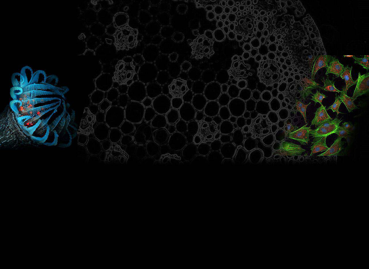
Fluorescence imaging is a potent method that gives researchers unmatched precision when studying and seeing numerous biological structures and processes. Fluorescence imaging filters, which selectively transmit certain wavelengths of light while blocking others, are at the core of this technology. The identification and visualization of fluorescent signals generated by target molecules or structures are made possible by selective filtering.
How Fluorescence Imaging Filters Work
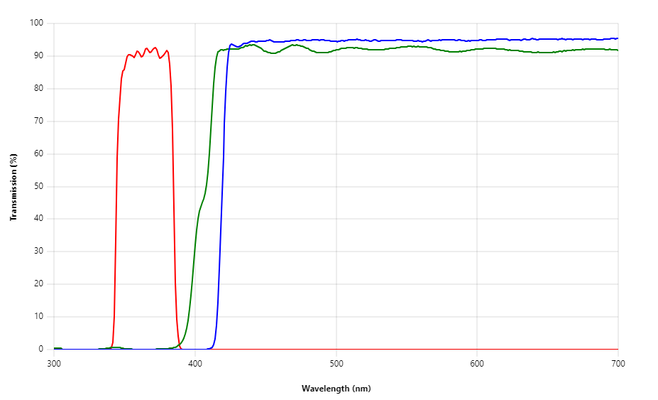
Multiple layers of specifically created materials with unique optical properties make up fluorescence imaging filters. These are coated on glass or other transparent substrates in a strategic manner. The excitation filter, dichroic mirror, and emission filter are the three main parts of a fluorescence imaging filter.
Excitation Filter: The first layer of the fluorescence imaging filter is the excitation filter. It transmits the excitation wavelength necessary to excite the targeted fluorophore or fluorescent molecule. Unwanted wavelengths are blocked by this filter, keeping them from reaching the sample.
Dichroic Mirror: The dichroic mirror is angled so that it reflects excitation light in the direction of the sample while letting emission light pass through. Making sure that only the desired fluorescence signals are picked up, it serves as a wavelength separator.
Emission Filter: The dichroic mirror is angled to reflect excitation light towards the sample while allowing emission light to pass through. Only the desired fluorescence signals are recognized since it serves as a wavelength divider.
The dichroic mirror is positioned at an angle, reflecting the excitation light in the direction of the sample while permitting the emission light to pass through. As a wavelength separator, it makes sure that only the desired fluorescence signals are picked up.
Advantages of Fluorescence Imaging Filters
The following benefits of fluorescence imaging filters make them essential tools in scientific research:
Enhanced Sensitivity: By selectively capturing the necessary fluorescence signals while reducing background noise, fluorescence imaging filters maximize the detection sensitivity. This makes it possible for researchers to clearly see even weak fluorescence signals.
Versatility: Researchers can adjust these filters to a variety of fluorescent markers and dyes by altering them to fit particular excitation and emission wavelengths. This adaptability makes it possible to examine various biological samples and their distinctive fluorescence properties.
Precise Localization: The exact localization of luminous molecules within biological samples is made possible by fluorescence imaging filters. Researchers can precisely determine the position of particular molecules by removing erroneous signals, revealing important details about cellular and molecular processes.

FAQs
Q1: Can fluorescence imaging filters be used in live-cell imaging?
Yes, live-cell imaging frequently employs fluorescence imaging filters. By recording the fluorescence signals released while reducing phototoxicity, they offer real-time imaging of dynamic cellular processes and interactions.
Q2: How do fluorescence imaging filters differ from other types of optical filters?
Fluorescence imaging filters, in contrast to conventional optical filters, selectively transmit and block specific wavelengths to improve the detection of fluorescence signals. Researchers can isolate and see particular components in complex biological samples because to this focused filtration.
Conclusion
In the world of scientific study and imaging methods, fluorescence imaging filters are a vital instrument. These filters improve the sensitivity and clarity of fluorescence signals by transmitting just the desired excitation and emission wavelengths. Because of their ability to localize precisely, researchers can examine and comprehend intricate biological processes at the cellular and molecular levels.
Fluorescence imaging filters give scientists a wide range of options for experimentation and discovery due to their adaptability and capacity to work with various fluorescent markers and dyes. These filters have changed a number of sectors, including biology, medicine, and materials research, by enabling improvements in fluorescence imaging techniques.
In conclusion, fluorescence imaging filters are essential for obtaining vivid and accurate fluorescence images. They greatly advance scientific research and facilitate ground-breaking discoveries thanks to their distinctive optical features and specific filtering abilities. Fluorescence imaging filters have the potential to help researchers better comprehend life and the natural world by helping them solve the mysteries of the tiny world.