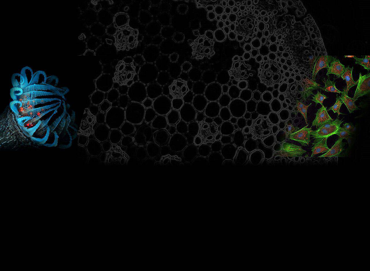
Fluorescence microscopy has revolutionized the study of cellular and molecular biology, enabling researchers to observe and analyze structures and processes within living cells with high sensitivity and precision. The key to this technique’s success lies in the use of fluorophores—molecules that absorb light at specific wavelengths and then emit light at longer wavelengths. The choice of fluorophore is critical for maximizing imaging efficiency, reducing photobleaching, and allowing for multiplexed detection of multiple targets simultaneously.
Among the vast array of fluorophores available, several stand out for their distinctive features and their ability to display bright, stable fluorescence. These include DAPI, AMCA (Aminomethylcoumarin), LysoSensor Blue, Alexa Fluor 350™, Marina Blue, Pacific Blue™, and sgBFP™. Each of these fluorophores has specific characteristics that make them suitable for particular applications, from live-cell imaging to subcellular localization studies. In this article, we will explore the unique properties of these fluorophores and their utility in modern fluorescence microscopy.
DAPI (4′,6-Diamidino-2-Phenylindole)
DAPI is a popular blue-emitting DNA stain, widely used for detecting and visualizing nuclei in fixed and live cells. It binds strongly to the minor groove of double-stranded DNA, making it an excellent choice for nuclear staining in fluorescence microscopy. DAPI’s fluorescence is typically observed under ultraviolet (UV) light, emitting at approximately 461 nm, which makes it ideal for imaging the nucleus in a variety of cell types. One of its most attractive features is its high fluorescence quantum yield, providing a bright signal even with relatively low concentrations. However, DAPI’s toxicity limits its use in live-cell imaging, making it primarily suited for fixed-cell applications.
Related reading:Fluorescence DAPI Optical Filters: Enhancing Imaging Accuracy and Sensitivity
AMCA (Aminomethylcoumarin Acetic Acid)
AMCA is a blue fluorophore with a peak emission around 446 nm. It is known for its high molar extinction coefficient and the resulting strong fluorescence signal, even at low concentrations. This makes AMCA a valuable tool for applications requiring high sensitivity, such as protein localization studies and cell viability assays. Its small size and water-solubility also make it suitable for conjugation to proteins, antibodies, and other biomolecules, enabling the tracking of specific molecules within living cells. Furthermore, AMCA is less prone to photobleaching compared to some other blue fluorophores, providing enhanced stability in prolonged imaging sessions.
LysoSensor Blue
LysoSensor Blue is a specialized fluorophore used for the detection of acidic organelles within the cell, particularly lysosomes. This pH-sensitive dye exhibits blue fluorescence when exposed to acidic environments, making it an invaluable tool for studying cellular processes related to endocytosis, lysosomal function, and autophagy. LysoSensor Blue is typically excited with ultraviolet light and emits fluorescence in the blue spectrum, providing clear visualization of acidic compartments within cells. By using LysoSensor Blue, researchers can gain insights into cellular metabolism and trafficking, as well as monitor changes in the acidic microenvironment associated with disease processes such as cancer.
Alexa Fluor 350™
Alexa Fluor 350™ is a blue-emitting, water-soluble dye with excellent photostability and minimal autofluorescence. With a peak emission around 440 nm, it is often used in multiplexed fluorescence imaging, where it can be paired with other dyes for multi-color experiments. One of its primary advantages is its high brightness and stability, even under intense laser excitation, which helps overcome issues of photobleaching that can limit the usefulness of other fluorophores. Alexa Fluor 350™ is commonly used for labeling proteins, antibodies, and other biomolecules in a variety of applications, including immunohistochemistry, flow cytometry, and live-cell imaging.
Marina Blue
Marina Blue is a bright blue fluorophore that exhibits fluorescence in the 450–470 nm range upon excitation. It is widely utilized in applications requiring high sensitivity and resolution, including single-molecule tracking, super-resolution imaging, and cellular imaging of small organelles or protein complexes. Marina Blue is particularly known for its ability to perform well in both fixed and live-cell imaging scenarios, and its stable fluorescence ensures reliable results over extended observation periods. Due to its high extinction coefficient and fluorescence quantum yield, Marina Blue is an ideal fluorophore for high-throughput screening assays.
Pacific Blue™
Pacific Blue™ is another popular blue-emitting fluorophore with a peak emission wavelength of around 455 nm. It is widely used for flow cytometry and immunofluorescence assays, where its sharp spectral profile and excellent brightness make it ideal for multi-color panel setups. Pacific Blue™ is designed to be highly photostable and resistant to fading, ensuring prolonged imaging sessions without significant signal loss. It is often used to label proteins, antibodies, and other molecules of interest, providing clear and distinguishable signals when combined with other fluorophores for multiplexed detection.
sgBFP™ (Super Green Fluorescent Protein)
sgBFP™ is a modified version of the classic blue fluorescent protein (BFP) and is engineered to be brighter and more stable than its predecessors. It emits in the blue range with a peak emission around 467 nm. sgBFP™ is particularly useful in genetic studies and live-cell imaging, where it can be used as a reporter protein to track gene expression and cellular dynamics. The increased brightness and photostability of sgBFP™ make it an ideal choice for long-term studies, such as monitoring dynamic processes in living cells, including protein interactions, gene expression, and cellular signaling.
Conclusion
The diverse range of fluorophores available today offers researchers a wealth of options for visualizing cellular structures and molecular interactions with unparalleled sensitivity and precision. Fluorophores such as DAPI, AMCA, LysoSensor Blue, Alexa Fluor 350™, Marina Blue, Pacific Blue™, and sgBFP™ each bring unique properties to the table, enabling tailored applications for a variety of microscopy techniques. The careful selection and optimization of these fluorophores based on their excitation and emission profiles, brightness, and stability are critical for ensuring high-quality results in fluorescence-based imaging experiments. As fluorescence microscopy continues to evolve, the ongoing development of new fluorophores and imaging technologies will undoubtedly further expand the possibilities for scientific discovery.
