Fluorescence microscopy technology can reveal the dynamic processes of cellular structures and biomolecules with extremely high resolution in modern biological research. In fluorescence microscopy, the selection and use of fluorescence microscopy filters enable clear, accurate images to be obtained.
This article will provide an in-depth discussion of filter technology in fluorescence microscopy, including various filter types, performance, and how to choose the appropriate filter for specific experimental needs.
What is Fluorescence Microscopy?
Fluorescence microscopy is a special type of optical microscopy designed to study the properties of organic and inorganic substances using fluorescence rather than relying solely on scattering, reflection, attenuation, or absorption. The technique typically employs a fluorescence filter cube containing three filters.
These filters facilitate the observation of different cells using different light sources and fluorophores, allowing for detailed cellular and molecular analysis. Fluorescence microscopy filters require appropriate blocking density OD values, high signal intensity, and high contrast.

What are Fluorescence Microscopy Filters
Fluorescence microscopy relies primarily on optical filters to select wavelengths, rather than using a monochromator. This type of microscope is usually configured with epifluorescence, which means that both the excitation and emission light passes through the same objective lens. Advantages of this configuration include:
- The excitation light is farther away from the detector: This helps reduce interference with the detector, improving image clarity and contrast.
- Co-location observation: Excitation and emission can be observed on the same optical path, facilitating simultaneous image capture.
- Partial reflection and scattering of excitation light: Although most of the excitation light passes through the sample, a considerable portion is reflected or scattered back to the objective lens, which helps enhance signal capture.
This design makes fluorescence microscopy particularly suitable for biological and medical research that requires high resolution and high contrast.

What are the filter types involved in the fluorescence filter cube?
The primary goal of fluorescence microscopy filter design is to create filter sets that maximize image contrast and maintain image quality to aid scientists, engineers, and researchers who use Optolong fluorescence filters.
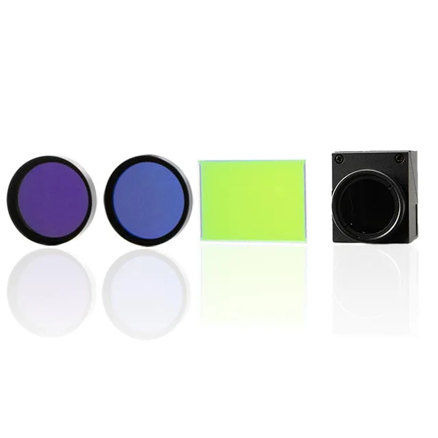
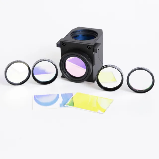
The main filter element in a fluorescence microscope is a set of three filters located in a fluorescence filter cube. They are an excitation filter, an emission filter, and a dichroic mirror beam splitter. Let’s discuss it together. What are the functions of these three filters in the fluorescence microscope filter set:
ExcitationFilter: An excitation filter is a high-quality optical glass filter commonly used in fluorescence microscopy and spectroscopy applications to select the excitation wavelength of source light. Most excitation filters select relatively short wavelengths of light from the excitation source because only these wavelengths carry enough energy to fully fluoresce the object being examined by the microscope. There are two main types of excitation filters used, short-pass filters and band-pass filters.
Emission filters: Also called barrier filters or emitters, attenuate all light transmitted by the excitation filter and very effectively transmit any fluorescence emitted by the sample while blocking unwanted traces of the excitation light. This light always has a longer wavelength (closer to red) than the excitation color. There are two main types of emission filters used, longpass filters and bandpass filters.
Dichroic Mirror: A dichroic mirror allows certain wavelengths of light to pass through, while other wavelengths of light are reflected. Dichroic beam splitters control which wavelengths of light enter their respective filters.
These three types of fluorescence filters are typically packaged together to form an excitation filter fluorescence microscope, an emission filter fluorescence microscope, and a dichroic mirror fluorescence microscope in a fluorescence cube so that the set can be inserted into the microscope together.

The Role of Fluorescence Filters
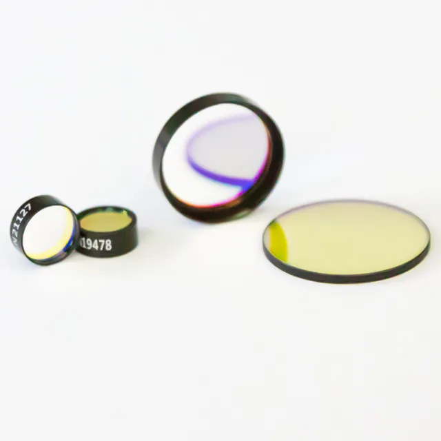
prolong Optics offers fluorescence filters that modify light within optical fluorescence imaging systems and are also used for observation purposes or to capture high-quality images using detectors. This delicate light management is not only related to image quality but also directly affects the accuracy and reliability of experimental data. They improve fluorescence imaging in several ways:
Increase contrast: Fluorescence filters work by selectively passing specific wavelengths of light (usually the fluorescent signal emitted by the sample) while effectively blocking other wavelengths of light. This selective delivery significantly increases the contrast of the image, making fluorescently labeled cellular structures or molecules more distinct from the background.
Block ambient light: There are often multiple light sources in a laboratory environment, such as indoor lighting and sunlight, which may interfere with observations under a microscope. Fluorescence filters can effectively block these unnecessary ambient lights, ensuring that the observation and imaging environment remains under ideal optical conditions.
Remove harmful ultraviolet or infrared rays: Ultraviolet and infrared rays may cause damage to samples, or cause photobleaching of certain fluorescent dyes, reducing their luminous efficiency. Using a fluorescence filter to filter out these harmful rays can protect the sample from damage while extending the life of fluorescent dye.
Selectively omitting or transmitting light of specific wavelengths: In fluorescence imaging, excitation light is usually required to excite the fluorescent molecules in the sample and make them emit light. Fluorescence filters selectively pass the wavelength of light used for excitation. At the same time, they block other unnecessary wavelengths to ensure that only the required light is absorbed and emitted by the sample.
Correct light path problems: Fluorescence filters can be used to correct certain defects in the optical system, such as chromatic aberration, which is the problem caused by different refraction angles of light of different wavelengths in the lens. With appropriate filters, the imaging quality can be improved to make the image clearer.
Reduce the intensity of light: In some cases, too strong light may cause saturation of the fluorescence signal, affecting image quality. Fluorescence filters can be used to appropriately reduce the light intensity entering the optical system to avoid signal oversaturation while protecting sensitive fluorescent molecules.
How to Choose the Best Filters for Fluorescence Microscopy?

Choosing the best filters for fluorescence microscopy is crucial for obtaining high-quality images and accurate data. The selection of filters can dramatically affect the clarity, contrast, and reliability of fluorescence signals. Here’s a step-by-step guide to help you select the most suitable filters for your fluorescence microscopy applications:
Determine the Fluorophores Used
Start by identifying the fluorophores you will use in your experiment. Each fluorophore has specific excitation and emission spectra.
Know the peak excitation and emission wavelengths of your fluorophores to match them with appropriate filters.
Match Filter Specifications to Fluorophore Properties
Check the bandwidth of the filters. Narrow bandwidth filters provide higher specificity and contrast but may reduce signal intensity. Wider bandwidth filters increase signal but can also increase background noise.
Ensure the filter’s transmission curve overlaps significantly with your fluorophore’s excitation and emission peaks for optimal performance.
Consider the Microscope Configuration
Verify compatibility with your microscope setup, including the light source and the optical components’ capabilities.
Some microscopes have fixed filter cubes, while others allow customizable configurations.
Assess the Application Needs
Consider the complexity of your sample. Multi-labeling experiments might require carefully selected filter sets to avoid bleed-through and cross-excitation. High-resolution applications may benefit from more precisely tuned filters to improve the signal-to-noise ratio.
Evaluate the Quality and Durability of Filters
Choose filters from reputable manufacturers to ensure durability and performance consistency. Consider filters with coatings that protect against degradation from UV exposure and general wear.
Budget and Cost-effectiveness
While higher-quality filters may be more expensive, they are generally more durable and provide better performance. Weigh the cost against the benefits in terms of improved image quality and reliability, so prepare your budget well before purchasing.
By carefully considering these factors, you can select the best filters for your fluorescence microscopy, enhancing your ability to explore and document biological phenomena at the microscopic level.
Conclusion
Fluorescence filters are core components for high-quality imaging. These filters not only enhance image contrast and clarity but also effectively manage light to protect samples and improve data accuracy.
To find the right optical filter for your application, please visit the OPTOLONG official website. They offer a variety of optical filters and provide related customization services. Contact us for an accurate quote!
