Introduction
Fluorescence microscopy has significantly advanced biological research by enabling highly precise visualisation of complex cellular components. The DAPI filter, which is an essential element of fluorescence microscopy, is critical in catching and detecting particular wavelengths produced by fluorophores. In-depth discussion of the wavelength range of DAPI filters, their uses, and how they support cutting-edge biological discoveries will be covered in this extensive essay.
What is the Wavelength Range of DAPI Filter?
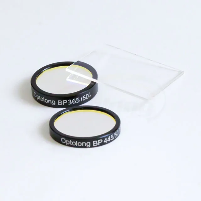
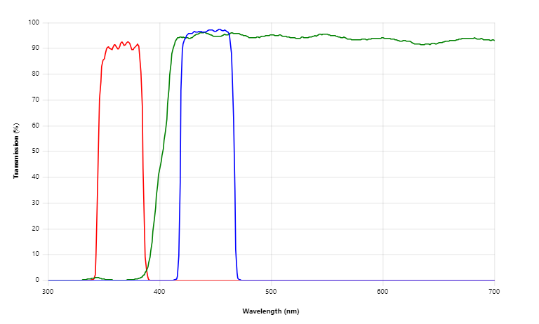
The ultraviolet (UV) spectrum forms the core of the wavelength range of DAPI filters. DAPI filters typically have a 440–460 nanometer emission range and an excitation range of roughly 340–360 nanometers. The DAPI fluorophore, a blue fluorescent dye that is frequently used to stain DNA in biological samples, is compatible only with these filters because of their particular design.
The Role of DAPI Filters in Fluorescence Microscopy
The principles of fluorescence, wherein particular molecules, known as fluorophores, produce light of a particular wavelength when activated by a light source, are the foundation of fluorescence microscopy. DAPI filters are crucial parts of fluorescence microscopes because they assist separate the excitation light from the fluorescent light that is emitted, producing images of the specimen that are crisper and more detailed.
Exploring DAPI Filter Applications
Cellular Nucleus Staining
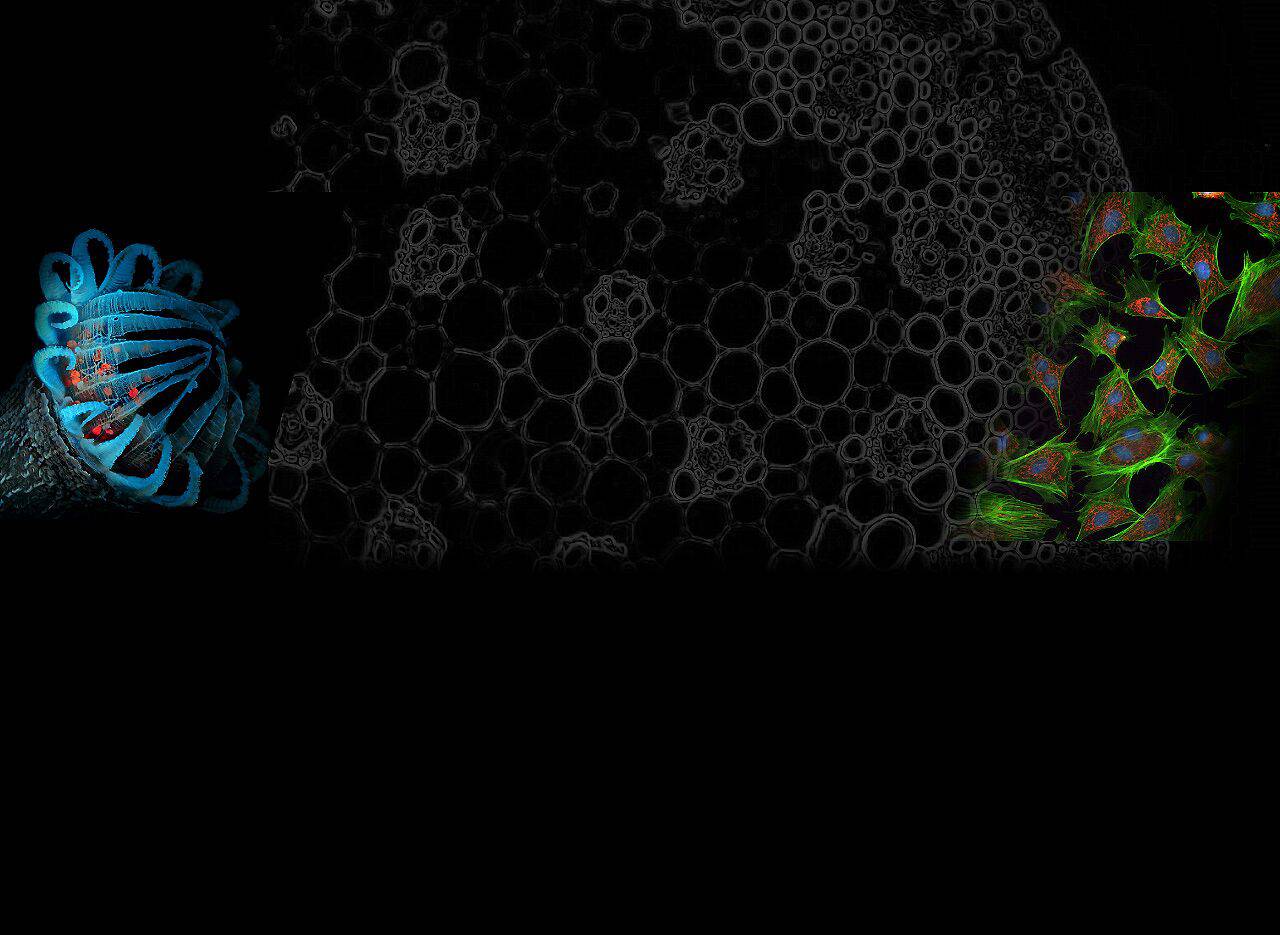
Staining cellular nuclei is one of the main uses for DAPI filters. Researchers can precisely visualise and map the distribution of genetic material within cells because DAPI has an affinity for DNA and attaches selectively to the nucleus of cells.
Cell Cycle Analysis
Cell cycle analysis use DAPI filters to more thoroughly comprehend how cells divide and enlarge. Labelling the DNA of cells with DAPI allows scientists to distinguish between the many cell cycle stages and get additional knowledge about a range of biological processes.
Fluorescence In Situ Hybridization (FISH)
FISH is an effective method for locating and detecting certain DNA sequences inside entire genomes or chromosomes. In FISH investigations, DAPI filters are frequently used to see the DNA target regions, assisting in genetic research and diagnostic applications.
Fluorescence Resonance Energy Transfer (FRET)
FRET is a sophisticated method for calculating molecular distances and interactions inside biological samples. To study protein-protein interactions and conformational changes in biological molecules, FRET experiments make use of DAPI filters together with other fluorescent labels.
Live-Cell Imaging
DAPI filters are essential for illuminating dynamic activities occurring within living cells during live-cell imaging research. Researchers can watch cellular activities in real-time and gain insightful knowledge about cellular behaviour by utilising DAPI in combination with other fluorescent probes.
Importance of DAPI Filters in Biological Research
DAPI filters are essential equipment in numerous biological research domains because to their adaptability and dependability. They have considerably advanced our knowledge of cellular functions, genetic processes, and disease causation.
FAQs about the Wavelength Range of DAPI Filter
Q: Why is the wavelength range of DAPI filters important?
Since it enables them to properly identify and record the distinct emission wavelengths of DAPI-stained samples, DAPI filters’ wavelength range is essential. Fluorescence images are improved in terms of quality and clarity thanks to DAPI filters, which separate the excitation light from the light that is emitted.
Q: Can DAPI filters be used with other fluorophores?
DAPI filters can be used with other fluorophores that produce light in a range close to that of the DAPI fluorophore, despite the fact that they are especially designed for the DAPI fluorophore. The spectrum overlap between various fluorophores must be kept to a minimum, nevertheless, in order to assure compatibility.
Q: How do DAPI filters improve image contrast?
By effectively blocking the excitation light and allowing only the released fluorescence to pass through, DAPI filters improve image contrast. This separation of the emitted light reduces background noise significantly, enhancing clarity and contrast in the final image.
Q: Are DAPI filters suitable for thick tissue sections?
Yes, thick tissue sections can be imaged using DAPI filters. The total image quality could be impacted by light scattering and absorption in thicker samples, though. In these situations, confocal or multi-photon microscopy can assist in overcoming these difficulties.
Q: Can DAPI filters work with fixed and live cells?
Yes, both fixed and live-cell imaging can be done with DAPI filters. Live cells can be stained with DAPI while they are still viable, but fixed cells are normally labelled with DAPI following fixation. The exact study goals will determine whether to use fixed or live-cell imaging.
Q: How do I choose the right DAPI filter for my experiment?
The microscope set-up, fluorophore compatibility, and the precise wavelengths of the excitation light source are a few of the variables that must be taken into consideration when choosing the right DAPI filter. Making the best decision may be aided by speaking with microscopy specialists and looking at the manufacturer’s specifications.

Conclusion
A key feature of fluorescence microscopy is the DAPI filters’ wavelength range, which enables scientists to see and examine cellular structures in incredibly fine detail. Scientists can analyse DNA distribution, look into cellular functions, and learn important new things about a wide range of biological phenomena by using DAPI filters. DAPI filters will undoubtedly stay crucial tools in every biologist’s toolbox as we continue to learn about the workings of life at the cellular level.