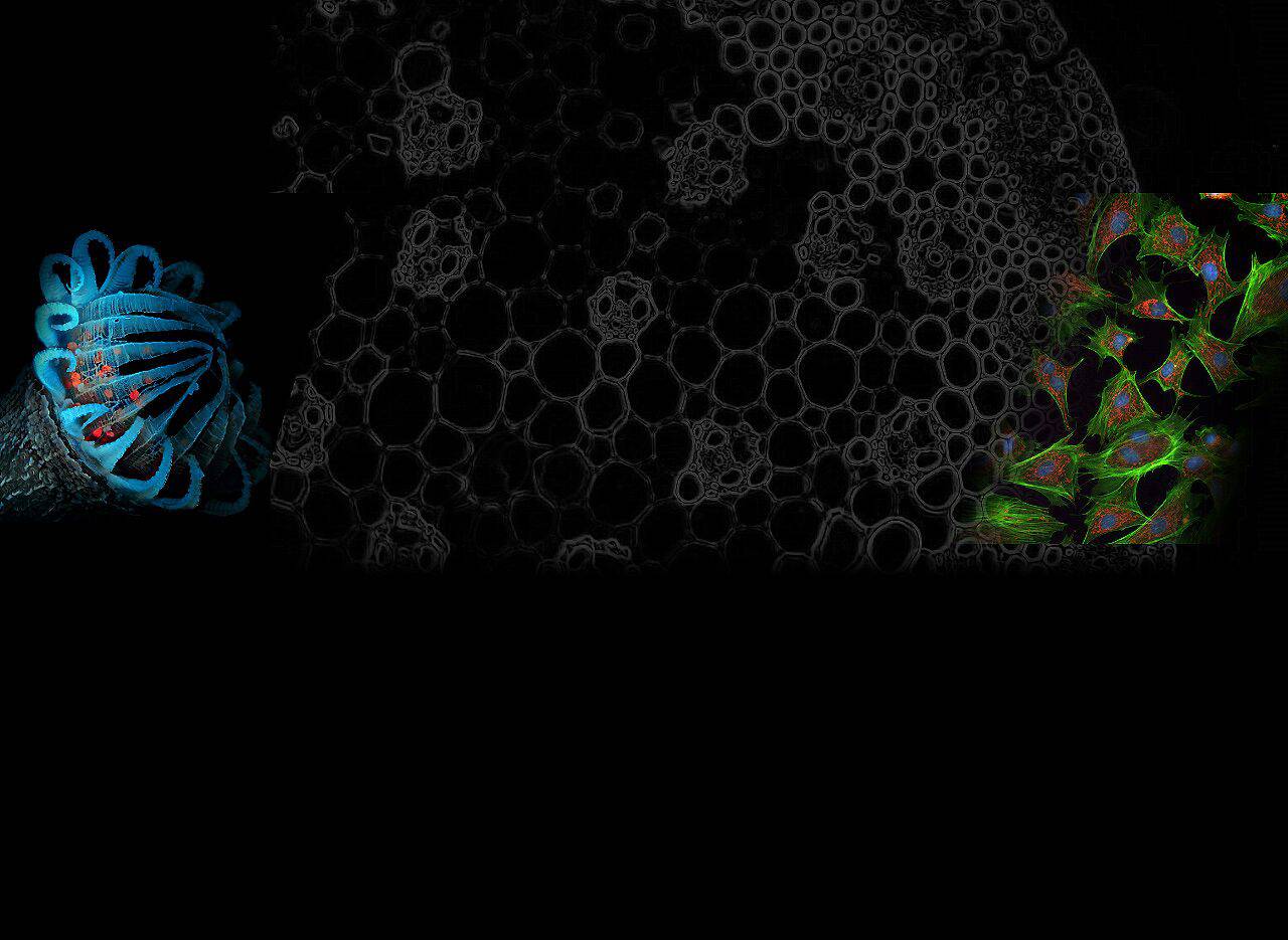
Fluorescence microscopes are widely used in fields such as biology, medicine, and material science to observe specific molecules in a sample that emit fluorescence. One of the key components of a fluorescence microscope is the excitation filter, which is responsible for selecting and filtering specific wavelengths of light used to excite fluorescent molecules in the sample.
Excitation filters precisely filter out extraneous spectral ranges to ensure that only the target fluorescent molecules are excited, resulting in high-resolution and high-contrast imaging. This article will explore excitation filters, how they work, and why they are useful for high-quality fluorescence microscopy.
What is an excitation filter?
An excitation filter is a type of optical filter that selectively allows specific wavelengths of light to pass through while blocking other wavelengths. In fluorescence microscopy, these filters ensure that only the light source’s appropriate wavelength reaches the specimen’s fluorophores.
When the fluorophore absorbs this light, it is excited and emits a longer wavelength of light, which can then be detected through the microscope.
How does it work?
In a fluorescence microscope, the light source typically emits a broad spectrum of wavelengths. The excitation filter is responsible for selecting a narrow range of wavelengths that correspond to the excitation peaks of the fluorophore being used. For example, if the fluorophore requires blue light (~450-480 nm) for excitation, the excitation filter will block all other wavelengths, transmitting only light in this range.
Once the excitation light reaches the fluorophore, it emits light of longer wavelengths, which passes through the emission filter and is detected by the camera or observer. The dichroic mirrors in the system help direct the excitation light to the sample while reflecting the emission light to the detector.
Importance in Fluorescence Microscopy
In fluorescence microscopy, choosing the right excitation filter is important for obtaining high-quality fluorescence images. The wrong excitation wavelength may result in the fluorophore not absorbing enough energy to fluoresce efficiently, or the emitted light may overlap with the excitation light, resulting in degraded image quality and weak signals.
Therefore, excitation filters should be selected based on the excitation and emission spectra of the fluorophore to minimize the spectral overlap between the excitation and emission light. Reducing this overlap reduces background noise and improves image contrast, resulting in sharper, higher-resolution images.
Advantages of excitation filters
1. Enhancement of fluorescent signal
The main advantage of using excitation filters is that they enhance the fluorescence signal by ensuring that the fluorophore absorbs a specific wavelength. This helps to increase the quantum yield, which is the ratio of absorbed photons to emitted photons, resulting in a more intense and stable fluorescence signal.
By maximizing the excitation efficiency of the fluorophore, the filter can provide a brighter image, making experimental results more reliable and reproducible.
2. Improved Specificity
The choice of excitation filters can significantly improve the specificity of an experiment. Each fluorophore is excited independently at a specific wavelength without interference from other fluorophores, allowing researchers to label and observe multiple different structures or molecules in the same sample. This multiplexing capability is especially important for complex experiments, providing multi-dimensional information without compromising image quality.
By precisely matching the excitation wavelength for each fluorophore, the excitation filter not only improves the sensitivity of the experiment but also avoids mutual interference between different fluorescent signals, enabling researchers to reliably detect multiple markers in the same sample, ensuring accurate and reproducible results.
3. Reduce background noise
Excitation filters significantly reduce background noise by blocking unwanted wavelengths that might otherwise interfere with or mask the fluorescent signal. By reducing background interference, excitation filters improve the signal-to-noise ratio, resulting in clearer images and more accurate data.
This is especially important for experiments that require high resolution and accurate results, ensuring that fluorescent signals stand out from the background and provide greater experimental reliability.
How to choose the right excitation filter?
When choosing an excitation filter, the excitation spectrum of the fluorophore should first be fully considered. Ideally, the filter should allow wavelengths near the absorption peak of the fluorophore to pass and block other irrelevant wavelengths as much as possible to maximize the intensity of the fluorescence signal and the clarity of the image.
Additionally, depending on the experimental needs, different types of filters can be selected, such as band-pass filters that transmit a specific range of wavelengths, and short-pass filters that block light longer than a specific wavelength.
Many fluorescence microscope systems also come with pre-configured filter blocks that integrate excitation filters, dichroic mirrors, and emission filters designed for specific fluorophores. This design facilitates rapid switching between different fluorescent dyes during an experiment, simplifying operations and increasing experimental flexibility and efficiency.
Conclusion
Excitation filters are a fundamental component of fluorescence microscopy, ensuring that the correct wavelength of light reaches the fluorophore and produces effective fluorescence emission. By choosing the right excitation filter, researchers can obtain high-quality, high-contrast images while minimizing background noise.
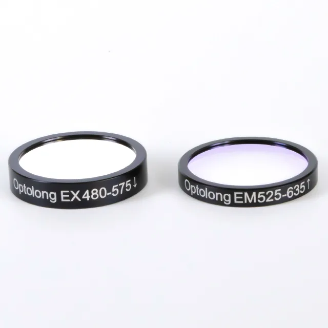
Optolong offers a wide range of high-quality optical filters designed for fluorescence microscopy and other scientific applications. Their filters include bandpass, shortpass, and custom optical filters designed to ensure precise wavelength selection for optimal imaging.
These filters improve signal-to-noise ratio, reduce background noise, and provide a sharper transition between the suppressed and transmitted regions. Optolong also offers pre-configured filter sets for specific fluorophores and life science applications, making them ideal for advanced imaging needs. Whether you are looking to purchase or customize a specific optical filter, please contact us for an accurate quote and information!
FAQ
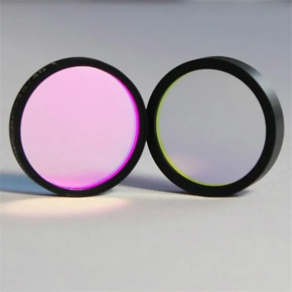
What is the difference between excitation and emission filters?
The main difference between excitation filters and emission filters is their function in the fluorescence microscopy examination process. Excitation filters selectively allow specific wavelengths of light to pass through, ensuring that only the light needed to excite the fluorophore reaches the sample. This maximizes excitation efficiency by blocking other unwanted wavelengths.
On the other hand, the emission filter is used after the fluorescence has been emitted from the sample. It allows only the fluorescent signal (typically at a wavelength longer than the excitation light) to pass through to the detector while blocking any remaining excitation light, ensuring that only the fluorescence is captured for imaging.
Do excitation filters degrade over time?
While physical filters are inherently robust, prolonged exposure to bright light can lead to photobleaching of the fluorophore rather than the filter. However, filters should be handled with care so as not to damage the coatings that control the light transmission properties.
What types of excitation filters are available?
There are generally two main types of excitation filters:
Short pass filters: Short pass filters allow light below a specific wavelength to pass through them.
Bandpass filters: Bandpass filters allow the transmission of a narrow range of wavelengths.
These filters are often used in conjunction with emission filters and dichroic mirrors in fluorescence microscopy.
Related reading: How dichroic mirrors split light?