Microscopes allow us to observe the tiny world in scientific research that the naked eye cannot see. Since their invention in the early 17th century, microscopes have played an important role in many fields such as biology, medicine, and materials science. A microscope is precisely designed and complex, consisting of multiple parts that work together to ensure that the device can accurately magnify and observe samples.
Understanding the functions of these parts helps us better use the microscope and also helps us understand the optical principles behind it. Next, we will look at the main parts that make up a microscope and how they work together to reveal the mysteries of the microscopic world.

A microscope is a complex instrument that consists of several key parts that can magnify and examine small objects. The following are the main components of a typical microscope:
- Eyepiece
- Objective lens
- Stage
- Stage clamp
- Light source
- Condenser
- Aperture or iris
- Coarse adjustment knob
- Fine adjustment knob
- Base
- Arm
Next, let us explain these microscope components one by one and clearly where they are located in the microscope and what their main functions are in the microscope.
Eyepieces
Mounted on the top of the microscope, this is the part through which the observer directly views the specimen. Eyepieces usually have a magnification of 10x or 15x and can be combined with the magnification of the objective to provide a higher total magnification.
Objectives
The main magnifying component on a microscope, usually with several objectives of different magnifications (such as 4x, 10x, 40x, 100x), is mounted on a rotatable nose wheel to quickly switch between different magnifications for observing specimens.
Stage
A platform for placing a slide with a specimen on it. The stage can usually be moved manually or automatically to allow precise positioning and observation of different parts of the specimen.
Stage Clamps
Clamps are fixed to the stage to stabilize the slide and prevent it from moving during observation.
Light Source
Provides light for observation, either from below or reflected light from above. Light sources are essential to illuminate the specimen and make it easier to observe.
Condenser
A lens located below the stage focuses light directly onto the specimen. Condensers are critical for improving the resolution and contrast of images.
Aperture or Iris
A device that adjusts the amount and quality of light hitting the specimen, is located below the condenser. By adjusting the aperture, the brightness and contrast of the image can be controlled.
Coarse Adjustment Knob
Used to quickly adjust the height of the stage (or objective) so that the specimen can be quickly brought into approximate focus.
Fine Adjustment Knob
Used to make finer focus adjustments to ensure a sharp image.
Base
The supporting structure of the microscope, to which all other parts are attached, either directly or indirectly.
Arm
Connects the base to the upper portion of the microscope (including the objectives and eyepieces), providing a grabbing point to facilitate moving the entire microscope.
What Role Do Optical Filters Play in Microscopes?

Optical filters in microscopes can significantly enhance the capabilities of a microscope. Here is a breakdown of the key roles optical filters play:
Enhancing Image Contrast and Quality
Color filters: Installed between the light source and the specimen, adjust the color of the light passing through the specimen, enhancing contrast or highlighting specific features of the specimen.
Neutral density filters: Reduce light intensity without changing the color, preventing overexposure and helping to achieve the desired balance of light in the view.
Selective Light Transmission
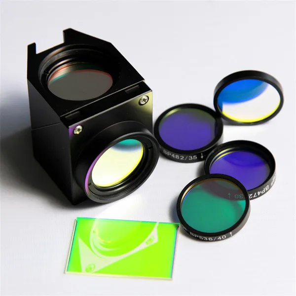
Bandpass filters: Allow only a specific range of light wavelengths to pass, which is particularly useful in fluorescence microscopy where specific wavelengths are required to excite fluorescent dyes.
Longpass and shortpass filters: Block shorter or longer wavelengths, respectively, which can help reduce unwanted light or enhance specific types of fluorescence.
Increasing Resolution
Polarizing filters are used to examine birefringent materials (such as crystals or stressed polymers) by filtering out light in specific planes. This not only improves image clarity but can also reveal stress distribution and molecular orientation.
Fluorescence Microscopy
Excitation and Emission Filters: In fluorescence microscopy, excitation filters allow only wavelengths that excite specific dyes to pass through, while emission filters block excitation light and allow only wavelengths emitted by fluorescent dyes to pass through. This separation enables clear, high-contrast fluorescence images.
Special Applications
Dichroic Mirrors (a type of filter): Reflect certain wavelengths while allowing others to pass through, helping to direct the path of light in complex microscopy setups such as confocal microscopes.
Heat-absorbing Filters: Protect sensitive specimens from heat damage, especially when using strong light sources such as lasers.
These filters are essential for researchers and professionals whose work depends on precise and clear microscopic images, ranging from biology and materials science to forensics and semiconductor manufacturing. By manipulating light in a variety of ways, optical filters enable more detailed and specific observations, making them an integral part of modern microscopy.
Where can I buy suitable filters for microscopes?
You can find filters for microscopes on the OPTOLONG website. They offer many types of optical accessories and support customized services. You can contact sales staff through their official website to get detailed product information and accurate quotes.
