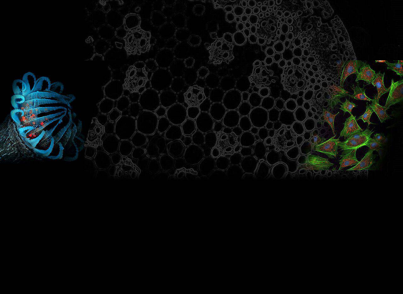
Fluorescence microscopy has revolutionized biological research by enabling visualization of cellular structures with high specificity and contrast. DAPI excitation emission plays a key role in this technique, providing a reliable method for DNA staining.
DAPI, or 4′,6-diamidino-2-phenylindole, binds tightly to adenine-thymine-rich regions of DNA. This binding results in a significant increase in fluorescence, making DAPI an invaluable tool for nuclear staining and chromosome analysis. Researchers frequently use DAPI in a variety of applications, including cell biology, medical diagnostics, and genetic testing.
What is DAPI Excitation and Emission?

DAPI (4′,6-diamidino-2-phenylindole) is a fluorescent dye specifically used for nucleic acid staining, especially for observing DNA under fluorescence microscopy. It works by tightly binding to adenine-thymine-rich regions of DNA.
- Excitation: Excitation of DAPI refers to the activation of the energy state produced by the dye after absorbing light of a specific wavelength (UV light). The optimal excitation wavelength of DAPI is around 359 nm.
- Emission: After excitation, DAPI releases energy and emits light, which is its emission process. The light emitted by DAPI is usually blue, with a spectral peak around 457 nm.
This excitation and emission process makes DAPI a very effective fluorescent marker for clearly showing the location and structure of the cell nucleus under fluorescence microscopy.
Optimization of Light Sources and Optical Components
Choosing the right light source will be more conducive to achieving optimal DAPI excitation emission. Modern fluorescence microscopes usually use high-intensity UV lamps or lasers to excite DAPI. These light sources provide the energy required to achieve maximum fluorescence intensity. Consistent and stable light output ensures reliable and reproducible results.
Filters and optical components can effectively enhance the DAPI excitation-emission process. Excitation filters selectively allow UV light to pass while blocking other wavelengths. Emission filters ensure that only blue fluorescence emitted by DAPI reaches the detector. High-quality optical components, including lenses and mirrors, ensure efficient light transmission and minimal signal loss.
The Optolong 30024 Single Band UV Filter Set DAPI is an example of a high-performance filter set designed for optimal DAPI staining. This filter set has excellent contrast and signal-to-noise ratio, making it suitable for both live and fixed cell imaging.

Scientific Research Results
- DAPI staining and its application in biological research: DAPI binds to DNA and emits blue fluorescence, making it widely used in techniques such as fluorescence microscopy, flow cytometry, and DNA staining assays.
- Comparison of DAPI and Hoechst dyes for nucleic acid detection: DAPI and Hoechst dyes differ in chemical structure, excitation/emission wavelengths, and binding affinity, but both are important for nucleic acid detection.
- Ideal nuclear counterstain: DAPI for fixed cells: DAPI is an ideal nuclear counterstain for fixed cells due to its relative brightness and enhanced fluorescence after binding to the AT region of dsDNA.
- Fluorescent dyes for DNA staining: DAPI can cross intact cell membranes to stain living cells, so it can be used in a variety of applications in biological research.
Application of DAPI in Fluorescence Microscopy
Cell Biology
The application of DAPI in fluorescence microscopy is extremely important, especially in cell biology and chromosome analysis. This dye can emit blue fluorescence under the excitation of ultraviolet light by specifically binding to the adenine-thymine-rich region of DNA, thereby achieving high-contrast imaging of the cell nucleus and chromosome structure.
This makes DAPI an important tool for studying cell cycle, nuclear morphology, and genetic organization and abnormalities, providing a reliable and effective method to observe and analyze the distribution and changes of nucleic acids in cells.
Medical Diagnosis
In the field of medical diagnosis, especially in cancer research and genetic testing, DAPI’s excitation and emission properties can specifically bind to DNA, and DAPI staining helps researchers identify and analyze nuclear abnormalities and chromatin structure in cancer cells, thereby gaining a deep understanding of the molecular mechanisms of cancer.
In addition, DAPI’s high specificity and bright blue fluorescence output make it an ideal tool for detecting subtle nuclear morphological changes associated with cancer.
In genetic testing, DAPI’s efficient DNA visualization function helps identify gene mutations and chromosomal abnormalities, providing reliable support for diagnosing genetic diseases and conducting prenatal screening. Researchers also often combine DAPI with other fluorescent probes for multi-parameter analysis to improve the accuracy and depth of diagnosis.
Comparison with Other Fluorescent Dyes

Specificity
DAPI excitation emission provides specificity for DNA staining. DAPI binds tightly to adenine-thymine-rich regions in double-stranded DNA (dsDNA).
This high specificity ensures that DAPI remains attached to DNA during various experimental procedures. The strong binding affinity produces clear and unique nuclear staining, making DAPI ideal for visualization of cell nuclei.
Sensitivity
DAPI excitation emission has excellent sensitivity. DAPI fluorescence increases approximately 20-fold upon binding to DNA. This high sensitivity enables researchers to detect even low concentrations of DNA. The bright blue fluorescence emitted by DAPI ensures high-contrast imaging, and this sensitivity can enhance detailed analysis by fluorescence microscopy.
Photobleaching
DAPI excitation emission faces the challenge of photobleaching. Prolonged exposure to strong UV light causes DAPI to lose fluorescence over time. This photobleaching effect reduces signal intensity and affects imaging quality. Researchers must optimize exposure times and use anti-fading reagents to mitigate photobleaching.
Toxicity
DAPI excitation emission has toxicity issues. DAPI can cross intact cell membranes and can therefore be used to stain living cells. However, DAPI can exhibit cytotoxic effects at higher concentrations, and researchers must carefully control DAPI concentrations to minimize potential toxicity, especially when working with live cells.
Summary
Understand the role of DAPI excitation and emission in fluorescence microscopy. DAPI binds tightly to DNA and fluoresces brightly in blue under UV light. This property makes DAPI a valuable resource for nuclear staining and chromosome analysis. Researchers benefit from the high specificity and sensitivity of DAPI. Future advances in fluorescence microscopy may enhance the applications of DAPI.
Innovations in filter technology, such as the Optolong 30024 Single Band UV Filter Set for DAPI, will improve imaging quality. Ongoing research will expand the usefulness of DAPI in the biological and medical fields.
If you want to purchase or consult about optical filters, please feel free to contact us. OPTOLONG provides various types of optical filters, including bandpass filters, dichroic mirrors, long-pass and short-pass filters, etc.
