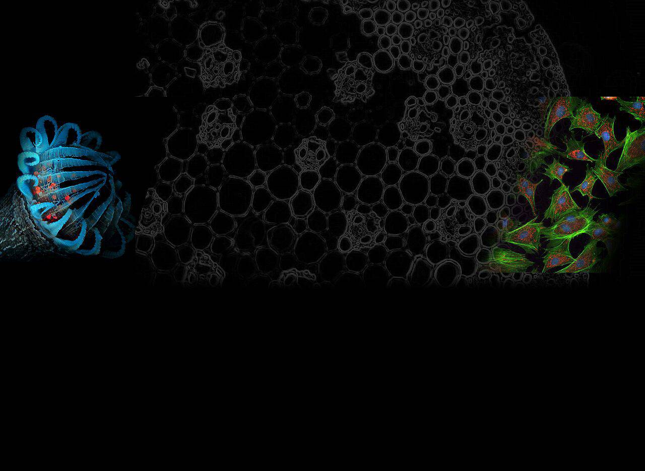
Fluorescence microscopy is a powerful technique used in scientific research to visualize and study fluorescently labeled samples.
The basics of fluorescence involve fluorophores absorbing light and then re-emitting it at a longer wavelength, creating a vibrant glow.
This phenomenon plays a crucial role in various fields such as cell biology, neurobiology, and immunology.
The importance of scientific research lies in its ability to provide detailed insights into cellular structures and processes. By labeling specific molecules with fluorescent dyes, researchers can track their movement and interactions within living cells, enabling the study of dynamic biological events.
Understanding how fluorescence microscopy works involves the utilization of fluorophores, which are molecules that absorb light energy and then emit it in a fluorescent form.
In fluorescence microscopy, excitation filters are used to selectively excite fluorescence, while emission filters are used to block the excitation light and allow only the fluorescence signal to pass through to obtain clear images and data.
How to Quantify Fluorescence
In scientific research, by quantifying the fluorescence emitted by fluorophores, researchers can measure the intensity of the fluorescent signal and provide valuable data for experiments.
When it comes to quantification, some basic principles can guide the quantification process. One common method is to measure the mean fluorescence intensity (MFI), which is the calculation of the average intensity of fluorescence in a given sample.
Additionally, researchers have utilized relative quantification to compare fluorescence intensity across samples or conditions, which allows for relative measurements without absolute quantification.
Fluorescence quantification not only provides accurate measurements, but also allows for comparisons between different experimental setups and conditions.
Steps to Quantify Fluorescence in Microscopy
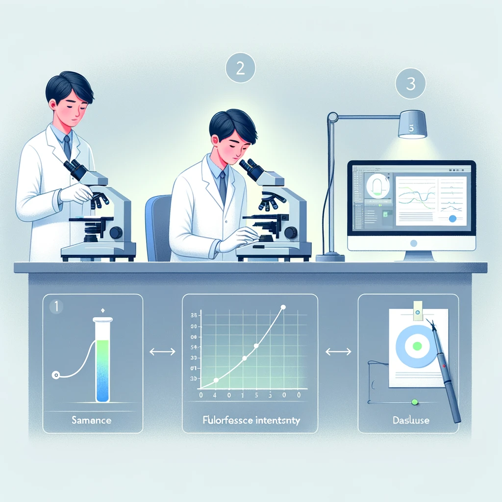
In the process of quantifying fluorescence in microscopy, several essential steps need to be followed to ensure accurate and reliable results.
Preparing Your Samples
When preparing samples for fluorescence microscopy, the first step is to select the appropriate fluorophore for the molecule or structure under study.
Different fluorophores have different excitation and emission spectra, so choosing the right fluorophore is critical to obtaining a clear and well-defined fluorescence signal.
In addition, the use of appropriate sample preparation techniques (e.g., immobilization, permeabilization, and blocking) helps to maintain the integrity of the sample while enhancing the binding of the fluorophore to the target molecule.
Capturing Images
Capturing high-quality images is fundamental to effectively quantifying fluorescence. Proper setup of the microscope includes optimizing parameters such as exposure time, gain, and resolution to ensure the best image quality without oversaturation.
Additionally, adherence to image capture best practices, including minimizing background noise and ensuring uniform illumination within the field of view, is also important for obtaining accurate fluorescence measurements.
Analyzing the Images
After capturing images of the fluorescent samples, the next step involves using image analysis software to process and analyze the data. This software allows for visualization, segmentation, and measurement of fluorescence signals within the images.
By applying appropriate algorithms and filters, researchers can accurately delineate regions of interest and quantify fluorescence intensity within these areas.
The software facilitates calculating fluorescence intensity, providing valuable quantitative data that can be further analyzed statistically.
Tools and Techniques for Quantification
1. Fluorescence Spectrometers
Fluorescence spectrometers are essential tools used to measure the fluorescence characteristics of samples.
They operate by emitting light of a specific wavelength onto the sample, causing the fluorophores to emit fluorescence, which is then detected and analyzed.
This process allows researchers to obtain valuable information about the excitation and emission spectra of fluorophores present in the sample.
How They Work
Fluorescence spectrometers work by directing excitation light onto the sample and measuring the emitted fluorescence.
The instrument then generates a spectrum representing the intensity of emitted light at different wavelengths, allowing researchers to analyze and quantify the fluorescent signals.
Applications in Quantification
These instruments find applications in various fields such as biochemistry, environmental science, and material research.
Researchers use fluorescence spectrometers to quantify the concentration of fluorescent molecules, study molecular interactions, and assess the purity of compounds based on their fluorescent properties.
2. Image Analysis Software
Image analysis software is an indispensable tool for quantifying fluorescence in microscopy. It offers numerous features for processing and analyzing images obtained from fluorescence microscopy experiments.
Popular options such as ImageJ and FIJI provide powerful tools for image segmentation, intensity measurement, and statistical analysis.
Popular Options
- ImageJ: A versatile open-source software with a wide range of plugins for customized image analysis.
- FIJI: An enhanced version of ImageJ with additional functionalities specifically tailored for biological-image analysis.
Tips for Effective Use
When using image analysis software for quantification, it’s essential to optimize parameters such as thresholding, background correction, and region-of-interest selection.
Additionally, utilizing built-in algorithms or creating custom scripts can streamline the quantification process while ensuring accurate measurements.
Common Challenges and Solutions
1. Dealing with Background Fluorescence
When conducting fluorescence microscopy, one common challenge is the presence of background fluorescence, which can interfere with the accurate quantification of specific signals.
To minimize background noise, researchers can employ techniques such as background subtraction algorithms and utilizing appropriate controls.
By subtracting the background fluorescence from the overall signal, researchers can obtain a more precise measurement of the actual fluorescent intensity emitted by their samples.
2. Ensuring Reproducibility
Achieving reproducibility in fluorescence quantification requires standardizing procedures to maintain consistency across experiments.
Researchers should establish standardized protocols for sample preparation, image capture, and analysis to minimize variability.
Additionally, regular calibration and maintenance of equipment such as microscopes and spectrometers are essential to ensure accurate and reliable measurements over time.
By implementing these measures, researchers can enhance the reproducibility of their fluorescence quantification results.
Conclusion
Recap of Key Points
- Fluorescence microscopy is a valuable tool for visualizing and studying fluorescent-labeled samples in scientific research.
- Quantifying fluorescence intensity is essential for accurate analysis and comparison of results across different studies.
- The process of quantifying fluorescence involves preparing samples, capturing high-quality images, and analyzing the data using image analysis software.
The Importance of Accurate Quantification
In scientific research, accurate fluorescence quantification enables researchers to measure and compare the intensity of fluorescent signals, providing valuable information for understanding cellular processes and interactions.
Accurate quantification ensures the reproducibility and reliability of scientific discoveries, thus advancing progress in fields as diverse as cell biology, neurobiology, and immunology.
Related reading: How to Use Fluorophores for Molecular Imaging